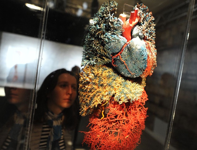
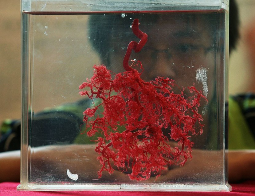
The main problem with bioprinted blood vessels was finally solved by a group of scientists.

Researchers at the University of Medical Center (UMC) Utrecht, Netherlands, worked together to make it possible. As of writing, health professionals are using bioprinted blood vessels for numerous medical purposes.
However, existing blood vessel bioprinting techniques usually rely on cell-friendly gels. These gels can result in structurally-inaccurate bioprinted blood vessels.
To solve this problem, UMC scientists combined melt electrowriting and bioprinting techniques to pursue functional blood vessels.
Problem With Bioprinted Blood Vessels Now Solved!
According to Interesting Engineering's latest report, UMC researchers used melt electrowriting because it is a highly accurate 3D printing method.

In order to achieve functional bioprinted blood vessels, UMC scientists used melt electrowriting to create a tubular scaffold.
After that, they submerged this scaffold into a vial with photoactive gel. Then, the submerged tubular scaffold was placed in the volumetric bioprinter.
Involved experts explained that the volumetric bioprinting method could be used to solidify cell-laden gels onto the scaffolds.
"In order to get this right, we had to place the scaffold exactly center in the vial," explained Gabriël Größbacher, the study's first author.
"Any deviation from the center would mean that the volumetric print would be offset. But we managed to center it perfectly by printing the scaffold on a mandril that we fitted to the vial," he added.
Study's Output
With the efforts of UMC scientists, they were able to print a proof of principle blood vessels with two layers of stem cells.
Aside from this, they also seeded epithelial cells in the center to cover the blood vessel's lumen.
But, Größbacher said that there's more to be done in their study. As of press time, the first author said that their next step is to bioprint an actual functional blood vessel.
If you want to learn more details about the new blood vessel bioprinting method that UMC scientists created, you can visit their official study, which was published at the Wiley Online Library.
© 2025 HNGN, All rights reserved. Do not reproduce without permission.


